|
JEPonline
Journal of
Exercise PhysiologyonlineISSN 1097-9751
An International Electronic
Journal for Exercise PhysiologistsVol 1 No 3 October 1998
Acute fluid volume changes in men during three days of creatine supplementation
Nutrition and ExerciseTIM N. ZIEGENFUSS1, LONNIE M. LOWERY2, AND PETER W.R. LEMON3
1Eastern Michigan University (Ypsilanti, MI), 2Kent State University (Kent, OH), and 3The University of Western Ontario (London, Ontario)
TIM N. ZIEGENFUSS, LONNIE M. LOWERY, AND PETER W.R. LEMON. Acute fluid volume changes in men during three days of creatine supplementation. JEPonline Vol. 1 No. 3 1998. Despite the plethora of recent research on creatine, little information exists relative to the nature of the weight gain reported from acute (< 5 d) supplementation. This study used multifrequency bioimpedance analysis (MBIA) to estimate the relative changes in total body water (TBW), extracellular (ECV), and intracellular (ICV) fluid volumes in 10 cross-trained and aerobically trained men before, and during three days of creatine monohydrate (Cr) supplementation (0.35 g · kg FFM-1· d-1). Cr ingestion produced clear trends in fluid shifts, and by day three had increased TBW 2% (0.86 ± 0.68 L, p = 0.07) and ICV 3% (0.77 ± 0.40, p < 0.01); no effect was noted in ECV (p = 0.51). These findings seem to indicate that the weight gain associated with acute Cr supplementation is primarily a result of water retention and that much of this increase is contained within the intracellular compartment.
Key words: ERGOGENIC AID, BIOIMPEDANCE, TOTAL BODY WATER, FLUID VOLUME
Introduction
A consistent finding in acute (i.e., 3-5 days) creatine supplementation studies is a weight gain of approximately 0.9-1.7 kg (1-3). Considering the rapidity of this response, most investigators have attributed it to increases in body water. This untested assumption is important because many supplement companies are promoting creatine (Cr) as a "research proven" way of increasing fat-free mass (FFM), implying these acute gains are composed of muscle protein. Interestingly, some investigators have shown that the distribution of body water between the intracellular and extracellular compartments can influence both protein and glycogen synthesis (4,5).Typically, measurements of fluid compartment volumes (e.g., total body water [TBW], extracellular fluid volume [ECV]) are made using tracer-dilution methods. This requires the ingestion or injection of a tracer solution (e.g., deuterium or tritium [TBW] and sodium bromide [ECV]), subsequent collection of blood, urine, or saliva sample(s) three to five hours later, and analysis using infrared spectroscopy or nuclear magnetic resonance (deuterium) and fluorescent excitation or high performance liquid chromatography (bromide). As noted by Johnson et al. (6) and Deurenberg and Schouten (7), these requirements make tracer-dilution methods tedious, time consuming, and sometimes variable.
The idea of using of multifrequency bioimpedance (MBIA) to assess fluid volumes is not new (8-10), and recent investigations have shown that MBIA can distinguish between, and assess changes in, the body fluid compartments of healthy humans (6,11-13). Briefly, MBIA is based on how the geometry of the body and its electrolytic composition affect the flow of a high frequency, low voltage electric current. The conductivity of FFM is greater than that of bone or adipose tissue because of its greater water (and therefore electrolyte) content. As a result, relatively small changes in the volume or conductivity of the body fluid compartments can significantly alter MBIA readings, allowing researchers to monitor fluid changes due to posture (13), pregnancy (12), and selective loss of ECV from diuretic drug use (7). Proponents of MBIA assert that at low frequencies (e.g., < 10 kHz) the capacitance of cell membranes prevents penetration of the electric current into the cell and therefore permits the estimation of extracellular fluid volume (10,14). In contrast, at high frequencies (e.g., > 50 kHz) the signal easily penetrates the membranes and passes through all fluids allowing the estimation of total body water (7). Although these assumptions are not without criticism, impedance values at low and high frequencies can be used to reliably estimate (via statistical regression) ECV and TBW, respectively (15). By subtraction then (TBW - ECV), ICV can also be estimated.
The aim of the present study was to estimate the relative changes in fluid volumes (TBW, ECV, and ICV) using the equations of Deurenberg et al. (11) during three days of Cr supplementation in cross-trained and aerobically trained men. Based on pilot data, it was hypothesized that Cr would increase TBW and ICV.
Methods
Subjects
Following Human Subject Review Board approval, ten healthy men were recruited from the student population at Kent State University to participate in the study. Seven of the subjects regularly (i.e., 3-4 d/wk, ~ 1 h/session) participated in a variety of sports (e.g., racquetball, roller blading, soccer, weight training) while the remaining three were competitive runners (i.e., 5-6 d/wk, ~ 100 miles/week). None of the subjects reported the use of tobacco, alcohol, or other medications and all regularly consumed meat in their diet. All reported to the laboratory after following standardized procedures (e.g., same time of day [morning, ± 30 min], > ten hours postprandial, > 24 hours postexercise). Prior to testing, written informed consent was obtained in from each subject; no monetary compensation was provided for their participation.Experimental Design
This experiment used a repeated measures design where each subject served as their own control. Specifically, subjects participated in six laboratory sessions. During the first three sessions, average baseline data (FFM, % body fat, TBW, ECV, and ICV) were obtained using MBIA. Sessions four, five, and six corresponded to 24, 48, and 72 hours of Cr loading, respectively. Strict dietary control was observed because changes in nutrition and hydration status could confound estimated fluid volumes. Specifically, during sessions one, two, and three, subjects completed detailed dietary records of all ingested foods. This included documentation of the quantity of food (estimated using dietetic measuring instruments provided by a dietician), method of preparation, and time of day ingested. Using these records, subjects were able to reproduce their diets during sessions four, five, and six (i.e., Cr loading period).Supplementation
All subjects were given a three day quantity of pharmaceutically supplied creatine monohydrate (American Biorganics, Aurora, OH) mixed into a container of powdered grape drink. This dosage plan was adopted in light of data suggesting the greatest increase in total intramuscular creatine (16) and PCr/ATP (17) resulting from endogenous supplementation occurs within 48-72 hours and that coingestion of creatine in a high glycemic beverage enhances uptake and deposition into skeletal muscle (18). The creatine dose was adjusted to FFM because differences in muscle mass affect the absolute size of an individual's creatine pool and have been suggested to contribute to large individual variations in the amount of creatine retained (1). Every three hours (with breakfast, lunch, dinner, and two snacks) subjects ingested 0.07 g · kg FFM-1 creatine monohydrate dissolved in 500 mL of grape drink (the powdered grape drink provided 34 g glucose per dose). This amount of creatine is approximately 10-20 times that found in a normal (mixed) diet and has previously been shown to elevate plasma [Cr] from 50-100 umol/liter to over 500 mmol/liter and increase total Cr and intramuscular PCr stores by up to 30% (1,3,16). For a 70 kg individual with 15% body fat, this dosage scheme amounts to approximately 21 grams per day.Bioimpedance Analysis
Following the measurement of each subject's stature (nearest 0.1 cm) and body mass (nearest 0.03 kg), resistance (R), reactance, impedance (Z) and phase angle were measured by a Xitron 4000 Bioimpedance AnalyzerTM (Xitron Technologies Inc., San Diego, CA) during a logarithmic sweep of frequencies ranging from 1-500 kHz. Diagnostic Lectec MP-3000 Tracet electrodesTM (LECTEC Corp., Minnetonka, MN) with a surface area of 700 mm2 were placed on the subject in a tetrapolar configuration (9). Specifically, current-detecting electrodes were placed between the styloid processes of the right radius and ulna, and between the medial and lateral malleoli of the right ankle. Current-introducing electrodes were then placed on the respective dorsal surfaces of the metacarpals and metatarsals, exactly five cm distal to the proximal (current-detecting) electrodes. Exact duplication of electrode placement was possible by marking contact areas with an indelible pen during session one. Electrode application was preceded by a light towel buffing to produce local hyperemia and a thorough scrubbing of the sites with alcohol swabs to remove dead skin and dirt.National Institutes of Health (NIH) standardization procedures for bioimpedance measurements were observed and followed (19): 1) all measurements were performed on a nonconductive table in an environmental chamber (23 degrees C) with the arms and legs slightly abducted (~ 30 degrees) from the trunk, 2) subjects were fasted (overnight) and had abstained from alcohol, caffeine, and heavy exercise for 24 hours, and 3) a 20 minute supine equilibration period preceded each measurement. Using these precautions, the variability of repeated (10 trials) R and Z values was determined to be < 2 (± 0.7) ohms. The average of three (serial) Z values at 100 and 1 kHz was used to estimate TBW and ECV, respectively, using the equations of Deurenberg et al. (11). Specifically, fluid volumes were estimated as:
TBW (L) = (height2 / Z100 x 0.34573) + (weight x 0.17065) - (age x 0.11) + 9.35where height and weight are measured in meters and kilograms. These equations have respective r2 and SEE of 0.95, 1.7 L (TBW) and 0.87, 0.9 L (ECV). Intracellular water volume was estimated by subtraction (TBW-ECV).ECV (L) = (height2 / Z1 x 0.19528) + (weight x 0.06987) - (age x 0.02) + 2.3
Data Treatment and Analysis
The Statistica software package (StatSoft Inc, Tulsa, OK) was used to analyze all data. All values are expressed as mean ± SD. Data from the first three (control) sessions were averaged and compared to sessions four (day 1 Cr), five (day 2 Cr), and six (day 3 Cr) with a one-way, repeated measures ANOVA to determine the effects of Cr supplementation on TBW, ECV, and ICV. A Newman-Keuls post-hoc test was used to identify the location of mean differences and effect size calculations were employed to assess the magnitude of treatment effects. Because this study was performed under highly controlled conditions and the changes in fluid volumes were expected to be close to the measurement error, the "conventional" alpha level (p < 0.05) was deemed too conservative. Instead, a 90% level of confidence (p < 0.10) was chosen, a priori, to indicate statistical significance. Nonetheless, actual p-values are reported so that readers can make comparisons to their own choice of critical values.Results
Subject Descriptive Characteristics
As indicated in Table 1, the subjects in this study formed a homogeneous sample. Following the regression principle of population specificity, the subjects' descriptive characteristics were very similar to those from which the Duerenberg et al. (11) equation was derived. Specifically, differences were (relative to the validation subjects): age (-1.3 yr), body mass (-3.3 kg), stature (-0.06 m), body fat (-2.3%), TBW (-2.5 L), ECV (-0.09 L), and ICV (-1.6 L). Aside from a significant (p < 0.01) gain in body mass (1.0 ± 3.5 kg), no side effects were reported from the supplementation protocol. Subjects consumed an average of 3671 ± 248 kilocalories per day, with carbohydrate, protein, and fat comprising 64 ± 10%, 12 ± 5%, and 24 ± 10%, respectively. No statistical differences were noted between diets under the control or creatine conditions.Fluid Volumes
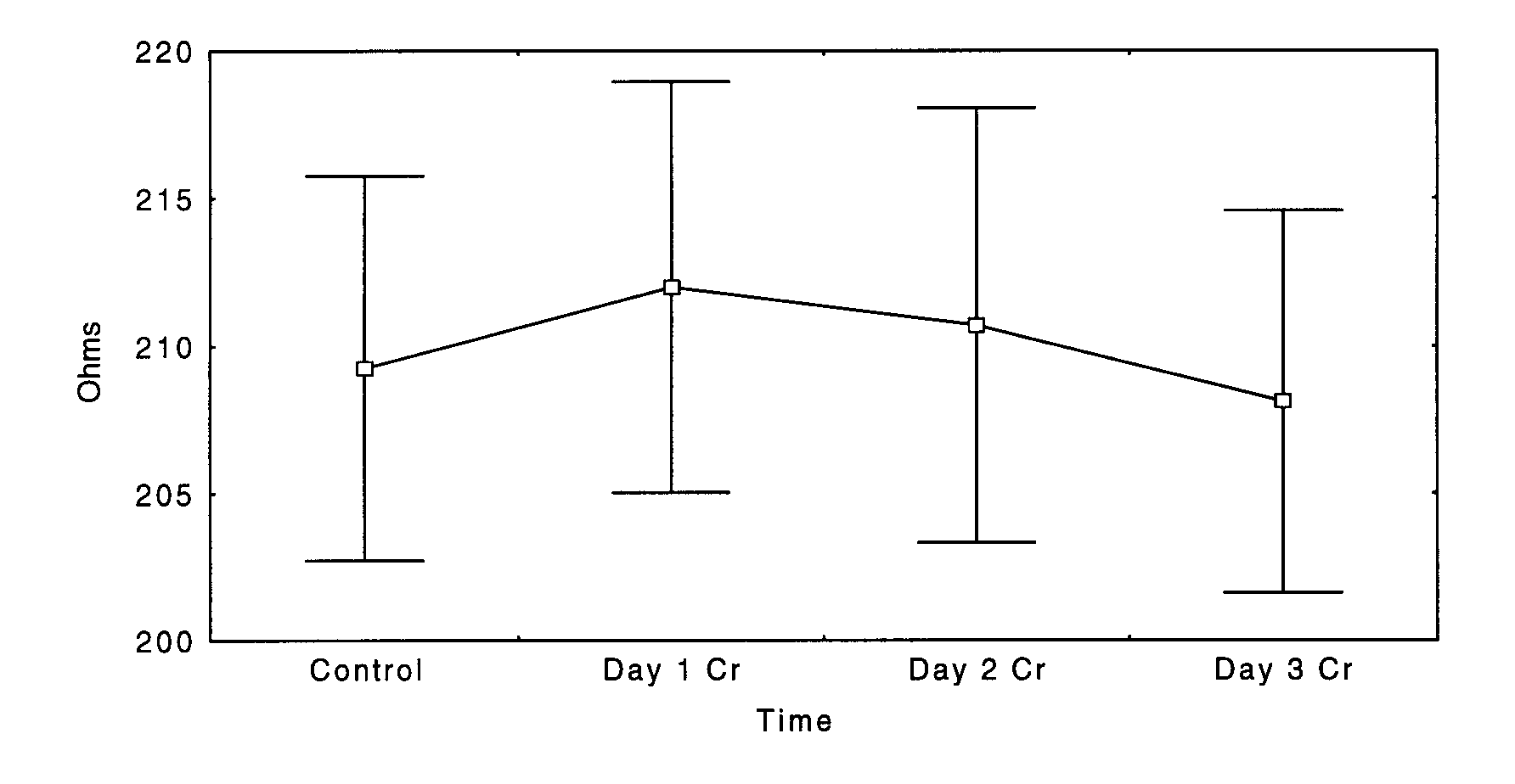
Estimated changes in TBW across the supplementation period are shown in Figure 1. A significant time effect was noted (p = 0.07) between the first and last days of Cr supplementation, where subjects gained 0.86 L of TBW. This represents a 2% increase in TBW.
Changes in ICV are shown in Figure 2. Statistically significant (p < 0.05) increases in ICV were verified between control and day 3 Cr (+0.57 L) and between day 1 and 3 Cr (+0.77 L). These increases represented gains in ICV of 2.3 and 3.1%, respectively. Fig 2. Mean ± SD changes in ICW (N = 10), * significantly greater than Control and Day 1 Cr (p < 0.05).
Changes in ECV were not statistically different throughout the supplementation period (p = 0.51). Changes in upper limb impedance paralleled whole-body changes, that is, increases in Z were found at lower frequencies (Figure 3) indicating reductions in extracellular fluid volume, while decreases were noted at higher frequencies (Figure 4) indicating increases in total limb water. Fig 3. Mean ± SD changes in upper limb impedance at 1 kHz (N = 10), * significantly greater than Control (p < 0.05).
Figure 4. Mean ± SD changes in upper limb impedance at 100 kHz (N = 10), observed values were not statistically different (p = 0.18).
Discussion
This study was undertaken to determine the effects of acute (i.e., three days) Cr supplementation on MBIA values associated with TBW (Z100) and ECV (Z1) and to estimate the relative changes in those fluid volumes using the equations of Deurenberg et al. (11). Although it is acknowledged the human body does not exactly fulfill the assumptions of MBIA (e.g., uniform cross-sectional area and homogenous conductivity), a 1994 review by the National Institutes of Health concluded that multifrequency bioimpedance is potentially just as accurate as fluid compartment assessments using labeled water (19). More importantly, the reproducibility of MBIA has been shown in several well-controlled investigations to surpass that of its criterion measure, tracer dilution (9,20). Specifically, over a wide range of age and body composition values, variations of 1-2% for successive R readings have been observed by Lukaski et al. (9) and Heitmann (20) among others. These same investigators reported intraclass correlation coefficients of 0.96-0.99 (as referenced in 20). Collectively, these factors support the use of MBIA to measure relative (within subject) changes in compartmental fluid volumes.In general, these results suggest that acute Cr ingestion elevates total body and intracellular fluid volumes but has no effect on ECV. It was anticipated that any increase in body mass would be explained largely by gains in TBW. These data indicate that increases in TBW account for approximately 90% of the acute gain in body mass. To our knowledge, this is the first study to report (relative) changes in fluid volumes during acute creatine supplementation in humans.
The small, yet statistically significant increases in estimated volumes of TBW and ICV highlight the compliant nature of the subjects involved in this study and our attempt to precisely control all factors that might confound the use of MBIA to estimate changes in fluid volumes (e.g. caffeine/alcohol use, diet and hydration status, and physical activity patterns, etc.). Further, our measurements were standardized according to the recent NIH conference statement to maximize our ability to detect a true difference when it existed. This is important because even small variations in the positioning and placement of electrodes, degree of limb abduction, ambient temperature, hydration status, meal ingestion, posture, length of time the subject is recumbent, and conductance of the examination can result in variable MBIA readings (19).
It has been suggested by Zillikens et al. (21) that whole-body bioimpedance measurements may not accurately predict changes in fluid compartments when changes are confined to specific body segments. Similarly, Baumgartner et al. (22) have demonstrated that whole-body R is determined primarily by the R of the extremities. Therefore, to verify the estimated changes in fluid compartment volumes, two additional analyses were performed. First, segmental (upper limb) impedance was measured. In all cases, upper limb Z values paralleled the changes found with whole-body MBIA, that is, increases in Z were found at 1 kHz while decreases were noted at 100 kHz. Moreover, upper limb ICV tended to increase while a (statistically) significant decrease was observed in (upper limb) ECV. Second, fluid volumes were recalculated using simple regression (i.e., only the impedance index term [height2 / Z]) to remove the possible confounding influence of changes in body mass. Again, statistically significant increases and decreases were noted in ICV and ECV, respectively. These important findings support the predictive value of the MBIA technique in estimating compartment specific fluid volume changes.
The mechanisms by which Cr supplementation increases TBW and shifts fluid into the intracellular space are unclear. It is known that Cr transfer from the extracellular space to the muscle sarcoplasm is governed by a Na+ dependent, saturable transporter residing in the plasma membrane (23), however, relatively little is known about the factors which regulate Cr uptake into skeletal muscle. One obvious factor regulating ICV is plasma osmotic pressure. Harris et al. (16) reported that plasma [Cr] peaks ~ 60 minutes following ingestion and steadily declines toward resting values over the next 5-8 hours. Considering MBIA values were obtained after an overnight fast, the possibility of residual plasma Cr affecting MBIA values, and therefore estimated fluid volumes, is unlikely. Theoretically, any change in the conductance of the MBIA current is caused by either a change in the composition of the conductor (i.e., water and electrolytes) or by a change in its length or diameter (24). Although plasma electrolytes were not measured, large differences are unlikely given the strict dietary control in this study. However, because 95-98% of Cr stores reside in skeletal muscle (1), it is possible that Cr supplementation results in an osmotic draw of fluid into the intracellular compartment (via increases in TCr and PCr), increasing ICV and TBW, ultimately increasing muscle volume. This explanation is consistent with these data as well as previous work from our laboratory demonstrating increases in muscle water and volume following Cr supplementation (17,25). These findings are exciting because of the potential for protein and glycogen synthesis to be enhanced via increases in ICV (4,5).
A useful way of estimating the degree of influence of the treatment (Cr supplementation) on the outcome (fluid volumes) is to calculate the standardized difference, effect size (difference between means / SD of the control group). This is particularly pertinent in exercise science where large differences between group means can be declared "nonsignificant" due to large variances and/or a small N (26). Cohen (27) and Thomas et al. (26) have suggested that effect sizes (ES) should be a primary product of research and should be analyzed in conjuction with p-values in order to offer valid standards of comparison with past and future investigations, independent of sample size. In general, ES of 0.2 may be considered small, 0.5 moderate, and 0.8 large (26). However, Hopkins (28) has recently computed that an ES as small as 0.20 (in competitive athletes) may be important because of its potential effects on performance. The calculated ES between control and day 3 revealed small effects of Cr supplementation on TBW (0.23) and moderate effects on ICV (0.48). ES values between day 1 and day 3 revealed stronger effects of Cr supplementation on TBW (0.39) and ICV (0.64). In summary, these data suggest that three days of Cr plus glucose supplementation in aerobic and cross-trained males increases MBIA estimated TBW and ICV, but has no effect on ECV. Future studies should verify these effects (and their magnitude) using more sensitive methods (e.g., magnetic resonance spectroscopy) and determine if these fluid changes are maintained with continued supplementation. If the latter is true, methods of body composition that assume a hydration constant (e.g., hydrostatic weighting) may incorrectly estimate changes in body fat and FFM during creatine supplementation. Finally, the influence of oral creatine supplementation on net protein status (protein synthesis minus protein breakdown) requires clarification as conflicting in vitro data have been reported (5, 29) and no in vivo human data are available.
References
1. Balsom P.D., K. Soderlund, and B. Ekblom. Creatine in humans with special reference to creatine supplementation. Sports Med 1994;18(4):268-280.
2. Earnest C., P. Snell, R. Rodriguez, A. Almada, and T. Mitchell. The effect of creatine monohydrate ingestion on anaerobic power indices, muscular strength and body composition. Acta Physiol Scand 1995;153:207-209.
3. Ziegenfuss T.N., D. Bredle, M. Rogers, B. Newcomer, M. Boska, and P. Lemon. Effect of creatine on repeated maximal muscle contraction. Can J Appl Physiol 1994;19:(suppl) 53.
4. Haussinger D., E.R. Roth, F. Lang, and W. Gerok. Cellular hydration state: an important determinant of protein catabolism in health and disease. Lancet 1993;341:1330-1332.
5. Ingwall J.S., C.D. Weiner, M.F. Morales, E. Davis, and F.E. Stockdale. Specificity of creatine in the control of muscle protein synthesis. J Cell Biol 1974;63:145-151.
6. Johnson H.L., P.S. Satinder, P. Mayclin, and T. Barbieri. Predicting total body water and extracellular fluid volumes from bioelectrical measurements of the human body. J Am Coll Nutr 1992;11(5):539-547.
7. Deurenberg P. and F. Schouten. Loss of total body water and extracellular water assessed by multifrequency impedance. Eur J Clin Nutr 1992;46:247-255.
8. Jenin P., J. Lenoir, C. Roullet, A.L. Thomasset, and H. Ducrot. Determination of body fluid compartments by electrical impedance measurements. Aviation Space Environ Med 1975;46:152-155.
9. Lukaski H.C., P.E. Johnson, W.W. Bolonchuk, and G.I. Lykken. Assessment of fat-free mass using bioelectrical impedance measurements of the human body. Am J Clin Nutr 1985;41:810-817.
10. Thomasset A. Electro-chemical volume measurements of extracellular liquids. Biophysics significance of impedance at one kilocycle. Lyon Med 1965;214:131-143.
11. Deurenberg P., A. Tagilabue, and F.J.M. Schouten. Multi-frequency impedance for the prediction of extracellular water and total body water. Br J Nutr 1995;73:349-358.
12. Lukaski H.C., W.A. Siders, E.J. Nielsen, and C.B. Hall. Total body water in pregnancy: assessment by using bioelectrical impedance. Am J Clin Nutr 1994;59:578-585.
13. Roos A.N., R.G.J. Westendorp, M. Frolich, and A.E. Meinders. Tetrapolar body impedance is influenced by body posture and plasma sodium concentration. Eur J Clin Nutr 1992;46:53-60.
14. Settle R.G., K.R. Foster, B.R. Epstein, and J.L. Mullen. Nutritional assessment: whole body impedance and body fluid compartments. Nutrition and Cancer 1980;2:72-80.
15. Van Loan M.D. and P.L. Mayclin. Use of multi-frequency bioelectrical impedance analysis for the estimation of extracellular fluid. Eur J Clin Nutr 1992;46:117-124.
16. Harris R.C., K. Soderlund, and E. Hultman. Elevation of creatine in resting and exercised muscle of normal subjects by creatine supplementation. Clin Sci 1992;83:367-374.
17. Lemon P., M. Boska, D. Bredle, M. Rogers, T. Ziegenfuss, and B. Newcomer. Effect of oral creatine supplementation on energetics during repeated maximal muscle contraction. Med Sci Sports Exerc 1995;27(5, suppl):S204.
18. Green A.L., E. Simpson, J. Littlewood, I. MacDonald, and P. Greenhaff. Carbohydrate ingestion augments creatine retention during creatine feedings in humans. Acta Physiol Scand 1996;158:195-202.
19. National Institutes of Health Technology Assessment Conference Statement. Bioelectrical Impedance Analysis in Body Composition Measurement (pp. 1-35), Bethesda, MD. 1994.
20. Heitmann B.L. Impedance: a valid method in assessment of body composition? Eur J Clin Nutr 1994;48:228-240.
21. Zillikens M.C., J. Willem, O. van den Berg, J. Wilson, and G. Roei-Swart. Whole-body and segmental bioelectrical-impedance analysis in patients with cirrhosis of the liver: changes after treatment of ascites. Am J Clin Nutr 1992;55:621-625.
22. Baumgartner R.N., Chumlea W.C., and A.F. Roche. Estimation of body composition from bioelectric impedance of body segments. Am J Clin Nutr 1989;50:221-226. 23. Guimbal C. and M. Kilimann. A Na+ dependent creatine transporter in rabbit brain, muscle, heart, and kidney. J Biol Chem 1993;268:8418-8421.
23. Guimbal C. and M. Kilimann. A Na+ dependent creatine transporter in rabbit brain, muscle, heart, and kidney. J Biol Chem 1993;268:8418-8421.
24. Hoffer E.C., C.K. Meador, and D.C. Simpson. Correlation of whole-body impedance with total body water volume. J Appl Physiol 1969;27:531-534.
25. Ziegenfuss, T.N. and P.W.R. Lemon. Three days of creatine supplementation improves anaerobic performance and increases muscle volume in high power athletes. (submitted)
26. Thomas J.R., W. Salazar, and D. Landers. What is missing in p < .05? Res Q Exerc Sport 1991;62(3):344-348.
27. Cohen J. Things I have learned so far. American Psychologist 1990;45:1304-1312.
28. Hopkins W.G. What is the smallest enhancement of competitive performance worth detecting in sport research? Med Sci Sports Exerc 1996;28(5, suppl):S187.
29. Fry, D.M. and M.F. Morales. A reexamination of the effects of creatine on muscle protein synthesis in tissue culture. J Cell Biol 1980;84:294-297.
Address for correspondence:Tim N. Ziegenfuss, Ph.D., 249 Warner Building, The Ruth Boughner Laboratory of Applied Physiology and and Clinical Assessment, Eastern Michigan University Ypsilanti, MI 48197 [phone: 734-487-1013, fax: 734-487-2024.
Acknowledgments.This project was supported in part by the National Strength and Conditioning Association and Quaker Oats. The authors gratefully acknowledge Wayne Sinning, Ed Freeman, Ron Mendel, and Phil Appicelli for their astute technical expertise.
Copyright ©1998
American Society of Exercise Physiologists
All Rights Reserved
ASEP Table of Contents